

Thin Cornea
🧠 Dr. Roque's Quick Answer
A thin cornea means the clear front window of the eye has less thickness than average. This matters in refractive surgery because laser procedures remove corneal tissue. Some people with a thin cornea can still have vision correction, but the decision depends on more than thickness alone. Corneal shape, stability, topography, prescription level, and overall eye health are just as important.
Many patients hear the phrase thin cornea during LASIK screening and immediately assume surgery is impossible. That is not always true. A thin cornea is an important safety finding, but it is only one part of a much larger decision. Some thin corneas are healthy and regular. Others are thin because they are structurally weak, irregular, scarred, or affected by conditions such as keratoconus. The key question is not simply, “Is the cornea thin?” The real question is, “Is this cornea safe for this particular procedure?”
In refractive surgery, safety comes first. A surgeon must protect the long-term strength and clarity of the cornea, not just reduce the eyeglass prescription. That is why thin cornea findings often trigger a more careful review of corneal imaging, tissue calculations, healing expectations, and alternative treatment options.
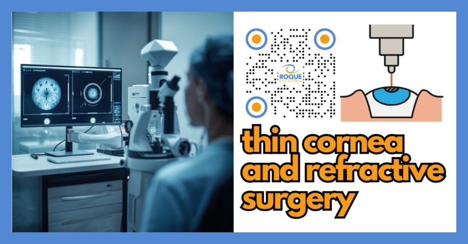
🧩 Focus: Thin cornea and its implications for refractive surgery
👁 Goal: Explain what a thin cornea means, why it matters, how it is evaluated, and which refractive options may or may not be suitable
🛡 Evidence-Based: Preferred Practice Patterns • Standards of Care • Systematic Reviews • Meta-Analyses
ROQUE REFRACTIVE SURGERY Knowledge Hub
Start with the complete guide:
🔬 Thin Cornea Anatomy Micro-Primer
- Cornea: The clear dome at the front of the eye. It provides much of the eye’s focusing power.
- Epithelium: The thin outer skin of the cornea. It heals quickly but does not provide the main structural strength.
- Stroma: The thick middle layer of the cornea. This is the main load-bearing part of the cornea and the layer most relevant to refractive surgery strength.
- Posterior cornea: The back surface of the cornea. Changes here can be an early clue that the cornea is not structurally normal.
📘 Thin Cornea Terminology Glossary
- Pachymetry: Measurement of corneal thickness.
- Topography: A map of the cornea’s front surface shape.
- Tomography: A 3D study of the cornea that helps assess front surface, back surface, and thickness distribution.
- Residual stromal bed: The remaining strong corneal tissue left after a laser procedure.
- Ectasia: Progressive corneal weakening and bulging that can occur in a vulnerable cornea.
- Keratoconus: A disorder in which the cornea becomes thin and cone-shaped, causing irregular vision.
Quick Navigation
Related Reading
Dr. Roque's Key Learning Points
- A thin cornea does not automatically mean all refractive surgery is impossible.
- Thickness alone is not enough. Corneal shape, symmetry, posterior surface, and tissue strength matter too.
- LASIK may be unsuitable in some thin corneas because it removes tissue and creates a flap.
- Some patients with thin but otherwise healthy corneas may still qualify for PRK, TransPRK, SMILE, ICL, or no surgery depending on the full work-up.
- The goal of screening is to avoid postoperative ectasia and protect long-term vision.
What a Thin Cornea Is
A thin cornea means the cornea measures less thickness than average on pachymetry or tomography. However, there is no single “magic number” that decides whether surgery is safe. A cornea may be thin but regular and stable. Another cornea may not look extremely thin, yet still be structurally unsafe because of abnormal shape or early ectatic disease.
This is why experienced refractive surgeons do not make decisions from thickness alone. They look at the entire structural picture: front and back corneal shape, thickness map, prescription to be treated, expected tissue removal, residual stromal bed, age, eye rubbing history, contact lens history, and signs of conditions such as keratoconus or forme fruste keratoconus.
💡 Dr. Roque's Analogy
Think of the cornea like the roof of a house. It is not enough to know the roof is thin. You also need to know whether the beams are straight, whether the shape is stable, and whether it can still support weight safely over time.
Why Thin Cornea Matters in Refractive Surgery
Laser refractive procedures reshape the cornea by removing tissue. In LASIK, tissue is affected by both the flap and the laser ablation. In PRK and TransPRK, there is no stromal flap, but tissue is still removed by the excimer laser. If too much tissue is removed from a cornea that is already thin or structurally vulnerable, the cornea may weaken over time and bulge forward. That complication is called corneal ectasia.
Corneal ectasia is one of the complications surgeons work hard to avoid during screening. That is why corneal topography and tomography are so important. They help identify irregular patterns, hidden weakness, abnormal thickness distribution, and early ectatic disease that may not be obvious during a standard eye exam.
Why a Cornea May Be Thin
Not all thin corneas are the same. Common possibilities include:
- Naturally thin but healthy cornea: Some people are simply born with thinner corneas.
- Keratoconus or early ectatic disease: The cornea becomes thinner and irregular over time.
- Previous corneal surgery: Prior refractive surgery can leave the cornea thinner.
- Corneal scarring or disease: Some conditions can distort thickness readings and alter structural safety.
- Contact lens warpage: Long-term contact lens wear can temporarily affect corneal measurements, which is why contact lens abstinence is often required before definitive imaging.
Tests Used During Screening
A good refractive surgery screening for thin cornea concerns usually includes several tests rather than one single measurement.
Pachymetry
This measures corneal thickness. It provides a number, but the number must be interpreted in context.
Corneal topography
This maps the front surface of the cornea and helps detect irregular astigmatism or suspicious shape changes.
Corneal tomography
This offers a more complete structural assessment. It evaluates front and back corneal surfaces and the thickness profile across the cornea.
Refraction and cycloplegic refraction
These help confirm the real prescription and determine how much corneal tissue would need to be treated if laser surgery is considered.
Ocular surface and tear film evaluation
Dry eye or surface disease can affect the accuracy of measurements and the quality of recovery.
Dilated retinal examination
This is especially important in myopic patients because retinal risks may coexist with corneal concerns.
Can You Still Be a Candidate for Vision Correction?
Possibly, but the answer depends on the full safety profile.
When LASIK may be unsuitable
If the cornea is too thin for the planned treatment, if the residual stromal bed would be too low, or if imaging suggests ectasia risk, LASIK may not be recommended.
When PRK or TransPRK may still be considered
Some patients with thin but regular corneas may still be candidates for surface ablation because no stromal flap is created. However, this is still a safety-driven decision. If the cornea looks suspicious or the treatment would remove too much tissue, surface ablation may also be inappropriate.
When SMILE may or may not fit
SMILE is another corneal laser option, but candidacy still depends on thickness, tissue profile, shape, and overall structural safety. A thin cornea does not automatically make SMILE safe.
When ICL may be a better option
Implantable collamer lens (ICL) surgery does not reshape the cornea. In some patients with a thin but otherwise healthy cornea, ICL may be more suitable because it preserves corneal tissue. That said, ICL has its own candidacy requirements involving anterior chamber depth, lens health, pressure considerations, and sizing measurements.
Alternative Options When the Cornea Is Too Thin for Laser Surgery
- Glasses: Still the safest and simplest option for many patients.
- Contact lenses: Soft toric lenses, rigid gas-permeable lenses, or scleral lenses may offer excellent vision depending on the corneal surface.
- ICL: A lens implanted inside the eye without removing corneal tissue.
- Cross-linking: In eyes with keratoconus or ectasia risk, corneal collagen cross-linking may be discussed to stabilize the cornea before any refractive plan is considered.
- No surgery for now: Sometimes the safest recommendation is observation and repeat imaging over time.
🚨 Dr. Roque's Emergency Warning
Urgent ophthalmic review is needed if you have rapidly worsening blur, increasing distortion, new glare with ghosting, sudden drop in vision, severe pain, or sudden redness after refractive surgery or after a corneal problem. These symptoms may signal ectasia, infection, inflammation, or another serious issue.
Main Risks and Concerns
- Postoperative ectasia in a structurally vulnerable cornea
- Undercorrection or overcorrection if the procedure choice is not ideal
- Dry eye symptoms and fluctuating vision
- Need for glasses or enhancement later on
- Progression of an unrecognized corneal disorder
Common Questions Patients Ask
Most patients want to know two things: “What is my safest option?” and “Can I still reduce my dependence on glasses?” A good surgeon answers both questions together. The right recommendation may be LASIK, PRK, SMILE, ICL, or no surgery. The correct answer depends on the whole eye, not just a single pachymetry number.
Why a “No” Can Protect Your Vision
Being told that your cornea is too thin for a certain laser procedure can feel disappointing, but it can also be a sign of excellent screening. The best refractive surgeons are not simply trying to make a patient qualify. They are trying to protect long-term corneal health and preserve quality vision for years to come.
Continue Reading
🏁 Dr. Roque's Take-Home Message
A thin cornea is an important refractive surgery finding, but it is not the whole story. Some thin corneas are safe for selected procedures, while others are not. The safest plan depends on careful screening, corneal imaging, tissue calculations, and honest discussion about alternatives. The goal is not just to reduce glasses—it is to protect long-term corneal strength and quality vision.
FAQ
1) Does a thin cornea automatically mean I cannot have LASIK?
No. A thin cornea raises an important safety concern, but candidacy depends on the full structural evaluation, not thickness alone. Some patients are ruled out for LASIK, while others may still qualify for another option.
2) What is considered a thin cornea?
There is no single thickness number that decides everything. Surgeons interpret thickness together with topography, tomography, prescription, residual stromal bed estimates, and overall corneal stability.
3) If I have a thin cornea, is PRK safer than LASIK?
Sometimes, but not always. PRK avoids a stromal flap, which may preserve more structural tissue than LASIK. However, if the cornea is abnormal or too vulnerable overall, PRK may still be unsafe.
4) Is ICL a good option for a thin cornea?
It can be. Because ICL does not remove corneal tissue, it may be attractive in some patients with thin but otherwise healthy corneas. It still requires a separate screening process for eye anatomy and lens suitability.
5) Can a thin cornea get worse over time?
A naturally thin but healthy cornea may remain stable. However, if the thinness is related to keratoconus or another ectatic disorder, the cornea can worsen over time. That is why imaging and follow-up matter.
6) Why do I need topography or tomography if pachymetry already shows the thickness?
Thickness is only one part of the story. Topography and tomography help reveal whether the cornea is regular, symmetrical, and structurally safe—or whether it has hidden patterns that increase risk.
📚 References
- American Academy of Ophthalmology. Refractive Surgery Preferred Practice Pattern®. Updated 2024.
- American Academy of Ophthalmology. Corneal Ectasia Preferred Practice Pattern®.
- American Academy of Ophthalmology. Corneal Topography.
- U.S. Food and Drug Administration. When is LASIK not for me?
- Raju RS, Raju CVG. Validation of an Artificial Intelligence-based Tool - The Screening Corneal Objective Risk of Ectasia Integrated into Anterion for Detection of Corneal Ectasia/Risk of Ectasia. Middle East Afr J Ophthalmol. 2024;30(4):257-265.
🤝 Roque Eye Clinic Patient Education Series
Dr. Manolette Roque | Dr. Barbara Roque
St. Luke's Medical Center Global City | Asian Hospital Medical Center
Philippines
Medical Review: Roque Advisory Council
Last Updated: March 2026
Medical Disclaimer
This article is intended for educational purposes only and does not replace professional medical consultation.


