

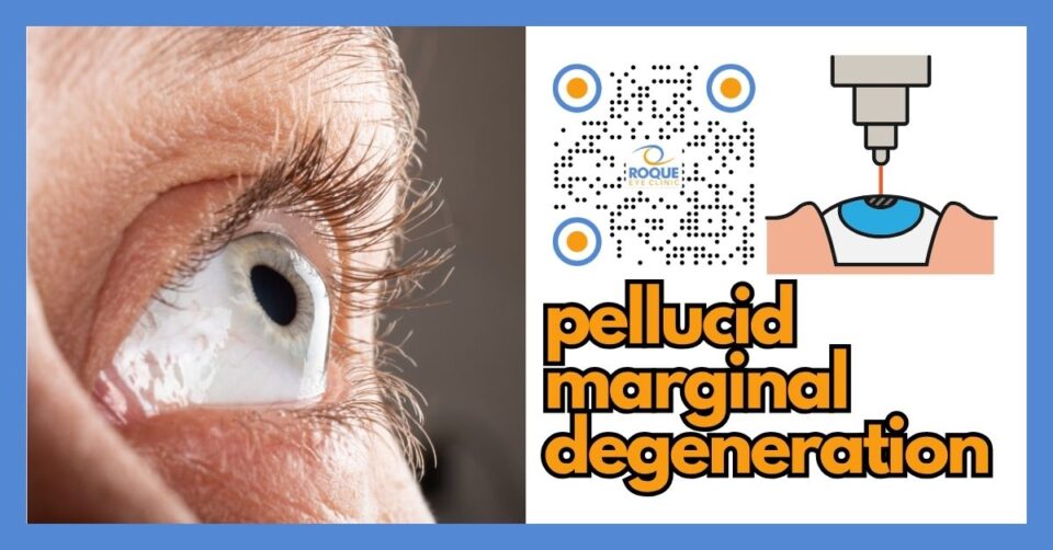
Pellucid Marginal Degeneration
🧠 Quick Answer
Pellucid marginal degeneration is a rare corneal ectasia in which the lower peripheral cornea becomes thin and causes irregular astigmatism and blurred vision. It can be mistaken for keratoconus, but recognizing it matters because laser refractive surgery such as LASIK or PRK may be unsafe. Many patients are managed with specialty contact lenses, while progressive or advanced cases may need cross-linking or corneal surgery.
Pellucid marginal degeneration, often shortened to PMD, is one of the most important corneal conditions to recognize during refractive surgery screening. It is uncommon, but it matters greatly because it can mimic other ectatic disorders, especially keratoconus, and because missing the diagnosis can lead to poor decisions about laser vision correction.
In plain language, PMD means that a lower band of the cornea becomes thinner than normal. The cornea then bulges just above that thinned zone, which creates irregular and often high astigmatism. Many patients first notice that their glasses no longer work well, that their prescription keeps changing, or that vision remains ghosted and distorted even after refraction.
🧩 Focus: Pellucid marginal degeneration as a corneal ectasia and refractive surgery contraindication
👁 Goal: Help patients understand what PMD is, how it affects vision, how it is diagnosed, and why it changes refractive surgery planning
🛡 Evidence-Based: Preferred Practice Patterns • Standards of Care • Systematic Reviews • Meta-Analyses
REFRACTIVE SURGERY Knowledge Hub
Start with the complete guide:
🔬 Pellucid Marginal Degeneration Anatomy Micro-Primer
- Cornea: The clear front window of the eye. PMD affects the shape and strength of this tissue.
- Peripheral inferior cornea: This is the lower outer part of the cornea. PMD classically causes thinning in this zone.
- Central cornea: In PMD, the center may look clearer and less obviously thin than the lower periphery, even though vision can still be badly distorted.
- Tear film and corneal surface: These help create a smooth optical surface. When the cornea becomes irregular, light no longer focuses cleanly and vision becomes blurred or warped.
📘 Pellucid Marginal Degeneration Terminology Glossary
- Corneal ectasia: Progressive thinning and bulging of the cornea that changes vision and weakens corneal shape.
- Against-the-rule astigmatism: A common astigmatism pattern in PMD in which the horizontal meridian is relatively steeper than expected.
- Topography: A map of the front corneal surface used to detect abnormal corneal shape.
- Tomography: A deeper structural map that analyzes both the front and back of the cornea and its thickness profile.
- Scleral lens: A large contact lens that vaults over the cornea and often improves vision in irregular corneas.
- Cross-linking: A treatment that strengthens corneal collagen and may help slow ectasia progression in selected cases.
Quick Navigation
Related Reading
Key Learning Points
- Pellucid marginal degeneration is a noninflammatory corneal ectasia with peripheral thinning, usually in the lower cornea.
- It often causes high and irregular astigmatism, so glasses may stop working well over time.
- PMD can look similar to keratoconus, so topography, tomography, and pachymetry are important.
- Recognizing PMD before refractive surgery is critical because LASIK, PRK, and similar corneal laser procedures are generally unsafe in ectatic corneas.
- Many patients are managed with specialty contact lenses; progressive or advanced cases may need cross-linking or corneal surgery.
What Pellucid Marginal Degeneration Is
Pellucid marginal degeneration is a clear, noninflammatory, peripheral corneal thinning disorder. The classic pattern is a crescent-shaped band of thinning in the inferior cornea, typically from about 4 o’clock to 8 o’clock, located just above the limbus. The cornea then protrudes above that band of thinning, producing irregular shape and worsening optical quality.
The word pellucid means clear. That is important because the cornea may remain clear and without obvious scarring or inflammation, even though its shape has become unstable. This can fool patients into thinking the problem is “just astigmatism,” when in fact the issue is a structural corneal ectasia.
💡 Analogy
Imagine a clear plastic bowl that becomes thin near its lower edge. The bowl may still look transparent, but once the wall weakens, the shape above that weak area starts to warp. PMD works in a similar way. The cornea may stay clear, yet its shape becomes distorted enough to blur and twist vision.
How PMD Differs From Keratoconus
PMD is often confused with keratoconus because both are ectatic corneal disorders and both can produce irregular astigmatism. The difference is mainly in where the thinning occurs and how the cornea deforms. Keratoconus usually involves more central or paracentral thinning and cone formation. PMD classically causes a lower peripheral band of thinning, with the protrusion occurring just above it.
On imaging, PMD may produce a classic butterfly, crab-claw, or kissing birds pattern, depending on the device and map being reviewed. This is why corneal imaging is so important. A slit-lamp exam alone may not be enough in early or subtle cases.
Symptoms and Visual Changes
Most patients notice a gradual decline in vision. Common complaints include:
- Blurred vision that glasses do not fully correct
- Frequent prescription changes
- Ghosting or double edges around letters
- Distorted shapes or stretched images
- Increasing astigmatism, often irregular
- Worse night vision or glare in some patients
Because PMD often progresses slowly over years, some people adapt without realizing how abnormal the vision has become. Others present only after repeated spectacle changes fail to restore good quality vision.
How Pellucid Marginal Degeneration Is Diagnosed
PMD is diagnosed through a combination of history, slit-lamp findings, refraction, and corneal imaging. A careful refractive surgery work-up often reveals the problem. Key parts of the evaluation may include:
1) Refraction and visual testing
Patients often show high astigmatism, sometimes irregular and difficult to refine. Vision may not fully improve with ordinary glasses. This is one clue that the cornea is not simply “strongly astigmatic” but structurally irregular.
2) Slit-lamp examination
The doctor looks for the characteristic peripheral band of thinning in the lower cornea. The cornea typically remains clear, avascular, and epithelialized, which helps distinguish PMD from inflammatory peripheral thinning disorders.
3) Topography and tomography
These are among the most important tests. They help detect the characteristic pattern of inferior steepening and irregular astigmatism, while also measuring front and back corneal shape and thickness distribution.
4) Pachymetry
Pachymetry measures corneal thickness. In PMD, the lower peripheral cornea may be abnormally thin, sometimes with a reversed thickness profile compared with a normal cornea.
5) Full refractive surgery screening
When PMD is being considered in a refractive surgery candidate, the evaluation should be complete. That means not only corneal mapping, but also careful refraction, corneal structural assessment, ocular surface review, and a dated eye health and fundus examination when indicated. The goal is to avoid missing a corneal ectasia before any permanent procedure is planned.
Why PMD Matters in Refractive Surgery
This is one of the most important sections for patients in a refractive surgery ecosystem. PMD is not just another source of astigmatism. It is a structural corneal weakness. That means corneal laser procedures such as LASIK and PRK can further weaken the cornea and may trigger or worsen ectasia.
In practical terms, PMD is generally treated as a contraindication to routine corneal refractive surgery. A patient with PMD may still be interested in vision correction, but the conversation changes completely. Instead of asking, “Can I have LASIK?” the safer question becomes, “How do we protect the cornea, improve vision, and avoid making the ectasia worse?”
This is why screening matters so much. A patient with unrecognized PMD who undergoes corneal laser ablation may have harmful outcomes. In contrast, a patient whose PMD is identified early can be counseled properly and guided toward safer management.
🚨 Emergency Warning
Seek urgent ophthalmic care if you have sudden severe pain, rapidly worsening blurred vision, a white or cloudy area on the cornea, sudden marked light sensitivity, or a feeling that vision has collapsed over hours to days. Advanced PMD can rarely be complicated by acute hydrops or even perforation, especially in very thin corneas or after trauma.
Who Gets PMD?
PMD is uncommon. It is often described in adults, frequently in young to middle adulthood, and many cases are bilateral, although the two eyes are not always equally affected. Some authors consider PMD part of a broader family of ectatic disorders related to keratoconus and keratoglobus. The exact cause remains uncertain.
Patients are not at fault for developing PMD. However, because ectatic disorders may worsen with chronic mechanical stress, many specialists advise minimizing frequent or forceful eye rubbing and protecting very thin corneas from trauma.
Treatment Options
Treatment depends on how much PMD is affecting vision, whether the cornea is progressing, and how advanced the thinning has become.
1) Glasses
In early disease, glasses may still help. Over time, though, many patients develop enough irregular astigmatism that spectacles no longer provide satisfactory vision.
2) Specialty contact lenses
This is often the main nonsurgical treatment. Rigid gas-permeable, hybrid, and especially scleral lenses can significantly improve optical quality by creating a smoother refractive surface in front of the irregular cornea. Lens fitting can be challenging because PMD affects the lower peripheral cornea, but many patients do very well with experienced fitting.
3) Corneal cross-linking
Cross-linking may be considered in selected progressive cases to strengthen corneal collagen and help slow further ectatic change. The exact role of cross-linking in PMD is still less standardized than in keratoconus, but it remains an important part of modern ectasia management discussions.
4) Intracorneal ring segments
Some selected patients may be considered for ring segments, but this is not routine for every PMD case. Very thin corneas can limit candidacy, and ring implantation may be contraindicated when thinning is extreme.
5) Corneal surgery
Advanced cases that no longer achieve useful vision with specialty lenses, or cases with severe structural distortion, may need corneal surgery. The exact surgical approach depends on the anatomy and surgeon judgment. Some patients require keratoplasty or other corneal reconstructive strategies.
Can PMD Be Cured?
There is no simple “cure” that restores a completely normal cornea. Instead, management focuses on three goals:
- Recognize the condition early
- Improve vision safely
- Reduce the chance of progression or structural complications
Many patients can function very well for years with the right visual rehabilitation and follow-up. The most dangerous mistake is not the diagnosis itself, but missing the diagnosis and treating the eye as if it were a normal astigmatic cornea.
Prognosis and Long-Term Follow-Up
PMD generally progresses slowly, often over years. That said, “slow” does not mean “harmless.” The irregular astigmatism can become visually significant, and advanced cases can be more difficult to manage. Lifelong follow-up is usually appropriate, whether the patient is wearing glasses, specialty lenses, or has undergone surgical treatment.
Patients should understand that even after intervention, they may still need glasses or contact lenses for best vision. The main goals are stability, safety, and quality of vision—not necessarily total spectacle independence.
What Patients Should Ask Their Eye Surgeon
- Do I truly have PMD, or another type of corneal ectasia?
- Which imaging tests show the diagnosis most clearly?
- Is my condition stable, or does it appear progressive?
- Are glasses enough, or do I need specialty contact lenses?
- Would cross-linking help in my case?
- Why is corneal laser refractive surgery unsafe for me?
- How often should I return for repeat imaging and follow-up?
Continue Reading
🏁 Take-Home Message
Pellucid marginal degeneration is a rare but important corneal ectasia that can cause severe irregular astigmatism and make routine refractive laser surgery unsafe. The key is early recognition, careful corneal imaging, and realistic management focused on protecting the cornea and improving vision safely. Many patients do well with specialty contact lenses, while progressive or advanced cases may need cross-linking or corneal surgery.
FAQ
1) Is pellucid marginal degeneration the same as keratoconus?
No. Both are corneal ectasias, but they are not identical. PMD usually causes peripheral inferior thinning with protrusion above that zone, while keratoconus more often causes central or paracentral cone-like thinning.
2) Can I still have LASIK if I have PMD?
In general, routine corneal refractive laser surgery such as LASIK or PRK is considered unsafe in PMD because the cornea is already structurally weak and at risk for worsening ectasia.
3) Why do my glasses stop helping as much?
PMD often creates irregular astigmatism. Glasses work best when the cornea is regular. As the shape becomes more distorted, spectacles may no longer provide clear, stable vision.
4) Are contact lenses useful in PMD?
Yes. Many patients achieve much better vision with rigid gas-permeable, hybrid, or scleral lenses because these lenses help neutralize the irregular corneal surface.
5) Does PMD always get worse?
Not always at the same speed, but PMD is generally considered a slowly progressive ectatic disorder. That is why periodic follow-up and repeat corneal imaging are important.
6) Can PMD cause an emergency?
Usually it progresses slowly, but advanced cases can rarely develop acute hydrops or even perforation, especially in very thin corneas or after trauma. Sudden pain or abrupt worsening of vision should be checked urgently.
📚 References
- EyeWiki. Pellucid Marginal Corneal Degeneration. Reviewed June 10, 2024.
- Sahu J, et al. Pellucid Marginal Corneal Degeneration. StatPearls Publishing. Updated 2023.
- Krachmer JH. Pellucid Marginal Corneal Degeneration. Arch Ophthalmol. 1978;96(7):1217-1221.
- American Academy of Ophthalmology. Corneal Ectasia Preferred Practice Pattern®. 2023, updated 2024.
- Tsatsos M, et al. Pellucid Marginal Degeneration: A Comprehensive Review of Diagnosis and Management. J Clin Med. 2025.
🤝 Roque Eye Clinic Patient Education Series
Dr. Manolette Roque | Dr. Barbara Roque
St. Luke's Medical Center Global City | Asian Hospital Medical Center
Philippines
Medical Review: Roque Advisory Council
Last Updated: March 2026
Medical Disclaimer
This article is intended for educational purposes only and does not replace professional medical consultation.

