

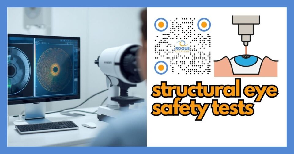
Structural Eye Safety Tests Before Refractive Surgery
🧠 Dr. Roque's Quick Answer
Structural eye safety tests before refractive surgery check whether your cornea and other eye structures are strong, regular, and healthy enough for laser vision correction or lens-based surgery. These tests look for hidden problems such as thin cornea, irregular corneal shape, keratoconus risk, abnormal healing risk, or other anatomical issues that can make surgery unsafe or less predictable.
Many patients think refractive surgery depends mostly on their eyeglass prescription. In reality, the shape, thickness, and structural stability of the eye matter just as much. A person may have a prescription that looks ideal for surgery, yet still be a poor candidate if the cornea is too thin, too irregular, or too vulnerable to weakening after treatment.
That is why structural eye safety testing is one of the most important parts of refractive surgery screening. These tests help your surgeon answer a basic safety question before anything permanent is done: Can this eye tolerate surgery safely?
🧩 Focus: Structural safety testing before refractive surgery
👁 Goal: Explain the tests that check corneal thickness, shape, stability, and other anatomical safety factors before laser or lens-based vision correction
🛡 Evidence-Based: Preferred Practice Patterns • Standards of Care • Systematic Reviews • Meta-Analyses
ROQUE REFRACTIVE SURGERY Knowledge Hub
Start with the complete guide:
🔬 Structural Eye Safety Tests Anatomy Micro-Primer
- Cornea: The clear front window of the eye. Laser vision correction reshapes this tissue, so its thickness and structural pattern matter greatly.
- Corneal stroma: This is the main support layer of the cornea. If too much tissue is weakened or removed, the cornea may become unstable.
- Anterior chamber: The space inside the front part of the eye. Its measurements matter more when lens-based procedures such as ICL are being considered.
- Pupil: The dark opening in the iris. Pupil size can affect glare, halos, and night vision symptoms after surgery.
📘 Structural Eye Safety Tests Terminology Glossary
- Pachymetry: Measurement of corneal thickness.
- Topography: A map of the front corneal surface.
- Tomography: A deeper 3-dimensional analysis of the cornea, including front and back surfaces and thickness profile.
- Keratoconus: A condition in which the cornea becomes thin and bulges forward, making laser surgery unsafe in many cases.
- Ectasia: Progressive corneal weakening and bulging that can happen in diseased corneas or rarely after refractive surgery.
- Biomechanics: The way corneal tissue bends, resists force, and maintains shape.
Quick Navigation
Related Reading
Dr. Roque's Key Learning Points
- Structural eye safety tests help determine whether your eye is anatomically safe for refractive surgery.
- These tests often focus on the cornea’s thickness, shape, back surface, and stability.
- A normal eyeglass prescription does not guarantee a safe cornea.
- Topography, tomography, and pachymetry are among the most important preoperative safety tests.
- Abnormal results may lead to a different procedure, more testing, postponement, or a decision not to operate.
Why Structural Eye Safety Tests Matter
Refractive surgery changes the way light focuses by altering the cornea or, in some cases, by placing or replacing a lens inside the eye. If the cornea is already weak, irregular, or suspicious for early ectatic disease, laser treatment can place that eye at higher risk. One of the most serious structural complications surgeons try to prevent is post-refractive ectasia, in which the cornea progressively weakens and bulges after surgery.
Good preoperative testing reduces the chance of missing subtle disease. In other words, these tests are not just paperwork. They are the structural safety net that protects patients from the wrong operation.
💡 Dr. Roque's Analogy
Planning refractive surgery without structural testing is like remodeling a roof after checking only the paint color. The support beams matter more than the surface look. In the eye, the “support beams” include the cornea’s thickness, shape, and strength.
Main Structural Eye Safety Tests Before Refractive Surgery
1) Corneal Topography
Corneal topography maps the front surface of the cornea. It helps detect irregular astigmatism, suspicious asymmetry, corneal steepening, and patterns that may suggest keratoconus or another corneal shape abnormality. This is one of the core tests in preoperative screening because a cornea can look clear at the slit lamp yet still show unsafe shape patterns on topography.
2) Corneal Tomography
Tomography goes deeper than topography. It analyzes the front and back corneal surfaces and the thickness profile across the cornea. This matters because early ectatic disease may show warning signs on the back surface or in the thickness distribution before obvious front-surface changes appear. In modern refractive screening, tomography is often one of the most valuable safety tests.
3) Pachymetry
Pachymetry measures corneal thickness. Surgeons use this information to estimate whether enough structurally safe tissue will remain after laser treatment. A thin cornea does not automatically mean “no surgery,” but it does mean the decision must be more cautious and individualized. In some cases, a different procedure may be safer than corneal laser surgery.
4) Corneal Biomechanical Assessment
Some clinics also use biomechanical testing devices that estimate how the cornea responds to force. These tests are not a replacement for topography or tomography, but they can add useful information when the risk profile is borderline. Biomechanics may help identify eyes that are more structurally vulnerable even if standard maps look only mildly suspicious.
5) Pupillometry
Pupil assessment is not mainly about structural strength, but it is still part of structural safety and visual-quality planning. A large pupil under dim light may increase the risk of glare, halos, and night-vision complaints if it is not considered during treatment planning and patient counseling.
6) Anterior Segment Imaging
Additional scans of the front of the eye may be used to assess corneal contour, epithelial thickness patterns, anterior chamber measurements, or other features important for procedure selection. These tests become especially relevant when ICL or another lens-based option is being considered.
What These Tests Try to Detect
- Thin cornea
- Irregular corneal shape
- Keratoconus or forme fruste keratoconus
- Pellucid marginal degeneration
- Suspicious posterior corneal elevation
- Abnormal thickness progression
- High ectasia risk
- Large pupil-related quality-of-vision concerns
- Anatomical mismatch for a planned procedure
What Surgeons Look for in the Results
Surgeons do not look at one number alone. They study the whole pattern. A cornea may be thick but irregular. Another may be thin but otherwise normal. Another may have a normal front map but suspicious back-surface elevation. Good screening combines multiple measurements, clinical judgment, and the patient’s age, refraction, contact lens history, ocular surface status, and family history.
That is why a patient should never assume that a quick “you qualify” statement based only on glasses power is enough. Safe screening is multi-layered.
Common Structural Red Flags Before Surgery
- Abnormal corneal asymmetry
- Inferior steepening or suspicious bow-tie patterns
- Unexplained thinning
- Posterior surface abnormalities
- Signs suggestive of early keratoconus
- Corneal scars that affect regularity or treatment planning
- Inadequate residual stromal safety margin for laser correction
- Poor agreement between different tests
🚨 Dr. Roque's Emergency Warning
If preoperative testing suggests progressive keratoconus, sudden corneal swelling, severe unexplained pain, or an acute eye problem, refractive surgery planning should stop until the underlying issue is properly evaluated and treated.
Why Contact Lens Discontinuation Matters Before These Tests
Soft, toric, rigid, and specialty contact lenses can temporarily change corneal shape. If testing is done too soon after contact lens wear, the maps can become misleading. A cornea may look irregular when it is only recovering from lens wear, or it may appear more regular than it truly is. That is why surgeons often require a contact lens holiday before structural testing.
Can a Patient Have Normal Vision but Unsafe Structural Tests?
Yes. This is one of the most important lessons in refractive screening. A patient may see well with glasses or contact lenses, have a refraction that looks treatable, and still have topographic or tomographic warning signs that make surgery unsafe. Structural tests help identify these hidden concerns before the cornea is permanently altered.
How Structural Test Results Affect Procedure Choice
These test results do not always lead to a simple yes or no. Sometimes they change the type of surgery being recommended. For example, a borderline cornea may steer the discussion away from LASIK and toward a flap-free option, or away from corneal laser treatment altogether and toward a lens-based procedure. In other situations, the safest answer is to avoid refractive surgery.
Structural testing can therefore lead to one of several outcomes:
- Proceed as planned
- Repeat tests after optimization or contact lens abstinence
- Choose a different refractive procedure
- Postpone surgery
- Avoid surgery because risk is too high
Why These Tests Matter Even More in Myopia
Many refractive surgery candidates are myopic, and myopic patients often seek correction at a younger age. Because myopia is so common, it can be easy to focus only on the prescription number. But structural testing is especially important here because ectasia risk detection, corneal thickness planning, and retinal screening all become very relevant in this group.
Questions Patients Should Ask About Structural Safety Tests
- Did my corneal maps look normal?
- Is my cornea thick enough for the procedure you are recommending?
- Did you see any sign of keratoconus or ectasia risk?
- Do I need to stop contact lenses longer and repeat the scans?
- Would another procedure be safer for my anatomy?
- Which structural finding is the most important in my case?
Continue Reading
🏁 Dr. Roque's Take-Home Message
Structural eye safety tests before refractive surgery are not optional extras. They are the foundation of safe planning. These tests help reveal whether the cornea is regular, thick enough, and stable enough for surgery. When the tests look abnormal, a careful surgeon may recommend more testing, a different procedure, or no surgery at all—and that caution can protect vision.
FAQ
1) What is the most important structural test before refractive surgery?
There is no single universal winner, but corneal topography, tomography, and pachymetry are among the most important tests because they help detect unsafe corneal shape and thickness patterns.
2) Can I still have surgery if my cornea is thin?
Sometimes yes, sometimes no. A thin cornea does not automatically rule out all procedures, but it does require more careful planning and may make some types of laser surgery unsafe.
3) Why do doctors worry about keratoconus before LASIK or PRK?
Keratoconus weakens and distorts the cornea. If it is missed before surgery, laser treatment can increase the risk of postoperative ectasia and poor visual outcomes.
4) What is the difference between topography and tomography?
Topography mainly maps the front corneal surface. Tomography gives a broader 3-dimensional picture, including front and back surfaces and thickness distribution.
5) Do contact lenses affect structural eye testing?
Yes. Contact lenses can temporarily change corneal shape, which is why patients are often asked to stop wearing them before scans.
6) Can these tests change the type of surgery recommended?
Yes. Structural findings may shift the plan from LASIK to another option, delay surgery, or lead to a recommendation not to operate.
📚 References
- American Academy of Ophthalmology. Refractive Surgery Preferred Practice Pattern®. Updated 2024.
- American Academy of Ophthalmology. Corneal Ectasia Preferred Practice Pattern®. Updated 2024.
- U.S. Food and Drug Administration. When is LASIK not for me?
- Randleman JB, et al. Risk assessment for ectasia after corneal refractive surgery. Ophthalmology. 2008;115(1):37-50.
- Raju RS, Raju CVG. Validation of the Screening Corneal Objective Risk of Ectasia for detection of corneal ectasia risk. Middle East Afr J Ophthalmol. 2024;30(4):257-265.
🤝 Roque Eye Clinic Patient Education Series
Dr. Manolette Roque | Dr. Barbara Roque
St. Luke's Medical Center Global City | Asian Hospital Medical Center
Philippines
Medical Review: Roque Advisory Council
Last Updated: March 2026
Medical Disclaimer
This article is intended for educational purposes only and does not replace professional medical consultation.

