

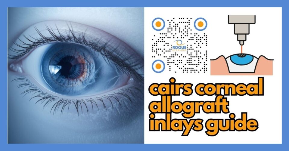
CAIRS Corneal Allograft Inlays
🧠 Quick Answer
CAIRS corneal allograft inlays are donor-corneal tissue segments placed inside the cornea to help regularize its shape, most often in keratoconus and related ectasia. They aim to improve vision quality and corneal symmetry without using synthetic plastic ring segments, but they are not suitable for every patient and still require careful screening, counseling, and follow-up.
CAIRS stands for corneal allogenic intrastromal ring segments. In simpler terms, this means specially prepared donor corneal tissue is inserted into channels within the patient’s cornea to help reshape and stabilize an irregular cornea. Although the article title uses the term “inlays,” many ophthalmologists discuss CAIRS as tissue ring segments placed within the corneal stroma rather than a classic central corneal inlay.
This procedure is mainly discussed in eyes with keratoconus or other corneal ectatic disorders, where the cornea becomes thinner, steeper, and more irregular. Instead of trying to remove tissue like laser refractive surgery, CAIRS is a tissue-addition strategy. That is one reason it attracts interest: rather than flattening the cornea by ablation, it changes corneal shape by adding biologic tissue in a controlled way.
🧩 Focus: CAIRS corneal allograft inlays for keratoconus and corneal ectasia
👁 Goal: Explain what CAIRS is, how it works, who may qualify, possible benefits, risks, recovery, and how it compares with other corneal options
🛡 Evidence-Based: Preferred Practice Patterns • Standards of Care • Systematic Reviews • Meta-Analyses
REFRACTIVE SURGERY Knowledge Hub
Start with the complete guide:
🔬 CAIRS Corneal Allograft Inlays Anatomy Micro-Primer
- Corneal epithelium: This is the thin outer skin of the cornea. It protects the eye and affects surface healing after surgery.
- Corneal stroma: This is the main structural layer of the cornea. CAIRS tissue segments are placed within this layer.
- Corneal cone: In keratoconus, part of the cornea bulges forward into a cone-like shape, causing irregular astigmatism and blurred vision.
- Visual axis: This is the central pathway of sight. Surgeons try to improve corneal shape while minimizing disturbance to the part of the cornea most important for clear vision.
📘 CAIRS Corneal Allograft Inlays Terminology Glossary
- CAIRS: Corneal allogenic intrastromal ring segments made from donor corneal tissue.
- Allograft: Tissue donated by another person of the same species, in this case human donor corneal tissue.
- Keratoconus: A disease in which the cornea thins and bulges, leading to irregular vision.
- Ectasia: Abnormal thinning and bulging of the cornea.
- Cross-linking: A procedure that uses riboflavin and ultraviolet light to stiffen and stabilize the cornea.
- ICRS: Intracorneal ring segments, usually synthetic ring implants used to reshape the cornea.
Quick Navigation
Related Reading
Key Learning Points
- CAIRS uses donor corneal tissue, not plastic, to help reshape an irregular cornea.
- It is usually discussed for keratoconus and corneal ectasia, especially when better corneal regularity is needed.
- CAIRS may be performed alone or together with corneal cross-linking in selected cases.
- The procedure aims to improve corneal shape and vision quality, but it does not guarantee freedom from glasses or contact lenses.
- Careful screening still matters because some eyes are better treated with cross-linking alone, specialty contact lenses, or corneal transplantation.
What CAIRS Corneal Allograft Inlays Are
CAIRS is a corneal tissue-addition procedure in which customized donor-corneal ring segments are inserted into channels created within the cornea. The goal is to make an irregular cornea more regular, reduce steepening, and improve how light enters the eye. It is most often discussed in patients with keratoconus, post-surgical ectasia, or similar corneal shape problems.
Older or more familiar ring-segment procedures often use synthetic intracorneal ring segments. CAIRS is different because it uses biologic donor tissue instead of synthetic plastic segments. This biologic approach is one of the main reasons many corneal specialists consider it interesting: donor tissue may behave more naturally within the cornea and may reduce some problems associated with synthetic materials, although it introduces its own considerations and is still not appropriate for every case.
💡 Analogy
If keratoconus makes the cornea behave like a dented or warped windshield, CAIRS is somewhat like adding carefully shaped support pieces within the windshield layers to help smooth and rebalance the surface. It does not create a brand-new cornea, but it may make the existing one work better.
How CAIRS Works
The surgeon first studies the patient’s corneal shape using topography and tomography. Donor corneal tissue is then prepared into segment shapes. Using a femtosecond laser or another precise method, channels are created in the patient’s cornea, and the donor segments are inserted into the stromal layer. These tissue segments change the way the cornea distributes its shape and curvature, often helping flatten steep areas and reduce irregularity.
In many real-world cases, CAIRS is discussed together with corneal cross-linking. The logic is straightforward: one part of the plan can help reshape the cornea, while the other can help stabilize it. Not every patient needs both, but the combination is common in progressive ectasia.
Why Surgeons Consider CAIRS
Patients with keratoconus often move through a ladder of treatment options. Early on, glasses or soft contact lenses may help. Later, rigid gas permeable lenses, hybrid lenses, or scleral lenses may provide better optical quality. If the cornea continues to distort or contact lens tolerance becomes poor, procedural options are considered. CAIRS belongs to that middle ground between contact lenses and full corneal transplantation.
That middle position is important. CAIRS is not usually the first treatment for mild, stable keratoconus, and it is not the same as a corneal transplant for advanced scarring. Instead, it may be considered when a surgeon believes the patient could benefit from corneal reshaping with tissue addition while preserving the patient’s own cornea.
Who May Be a Candidate for CAIRS
Potential candidates usually have keratoconus or another ectatic corneal disorder with irregular corneal shape affecting vision. They may have poor visual quality with glasses, contact lens intolerance, or corneal steepening that makes additional support and regularization attractive. Some patients may already have undergone other corneal treatments, while others may be evaluated for CAIRS before moving toward transplantation.
However, candidacy is not based on diagnosis alone. Your surgeon must review:
- Corneal thickness and shape
- Location and severity of the cone
- Presence of scarring
- Progression status
- Contact lens history and tolerance
- Visual goals and expectations
- Need for combined cross-linking
- Whether a transplant would be more appropriate
In other words, CAIRS is highly individualized. Two patients with “keratoconus” can have very different recommendations depending on thickness, scarring, cone position, age, progression, and contact lens performance.
Potential Benefits of CAIRS
1) Biologic tissue rather than synthetic material
One major attraction is that the implanted segments are made of donor corneal tissue. Many surgeons see this as a potentially more biocompatible approach than synthetic plastic ring segments.
2) Improvement in corneal regularity
CAIRS aims to make the corneal shape less irregular. When that happens, patients may notice better quality of vision, improved spectacle correction, or improved contact lens tolerance.
3) May delay or reduce the need for transplant in selected cases
For the right patient, CAIRS may serve as an intermediate step before corneal transplantation rather than an immediate jump to a bigger surgery.
4) Can be combined with cross-linking
When progression is an issue, surgeons may combine reshaping with stabilization in a staged or simultaneous plan.
Limitations and Trade-Offs
It is not a cure for keratoconus
CAIRS does not make keratoconus disappear. It is a structural and optical management strategy. Some patients still need glasses, specialty contact lenses, or later procedures.
It does not fit every cornea
Very advanced scarring, severe thinning, active disease issues, or anatomy that cannot support the planned channels may make CAIRS unsuitable.
Recovery and outcomes vary
Although many eyes improve, the amount of improvement differs from patient to patient. Some eyes gain meaningful visual quality, while others gain mainly topographic improvement or better contact lens tolerance rather than dramatic unaided vision.
Follow-up is still essential
Because the underlying ectatic disease still matters, continued monitoring remains important even after technically successful surgery.
Recovery and Follow-Up
Recovery depends on the exact surgical method, whether cross-linking is combined, ocular surface condition, and how the cornea heals. The first days may involve light sensitivity, foreign-body sensation, watering, blurred vision, or discomfort. Vision often remains variable during early healing.
Many patients need repeated visits so the surgeon can check the wound, segment position, surface healing, corneal clarity, and visual progress. If cross-linking was combined, recovery may be more noticeable than a ring-segment procedure alone. Like many corneal procedures, the cornea may keep remodeling for weeks to months.
🚨 Emergency Warning
Seek urgent ophthalmic review if you develop rapidly increasing pain, marked redness, discharge, sudden major drop in vision, a white corneal spot, or severe light sensitivity beyond the expected early postoperative course. These may signal infection, inflammation, melt, or another serious complication.
Risks and Complications to Discuss
No corneal procedure is risk-free. Reported or discussed risks with CAIRS may include:
- Infection
- Inflammation
- Delayed healing
- Segment position issues
- Insufficient visual improvement
- Residual irregular astigmatism
- Need for continued contact lens use
- Need for additional surgery
- Rare tissue-related complications such as melt or necrosis
These risks are one reason careful case selection matters. A procedure that is promising in published case series may still be the wrong choice for a specific patient sitting in clinic today.
CAIRS vs Synthetic Ring Segments
Both approaches aim to reshape an irregular cornea, but they do so with different materials. Synthetic ring segments use manufactured implants. CAIRS uses biologic donor corneal tissue. Potential advantages of CAIRS may include biocompatibility and a tissue-based approach. Synthetic segments, however, are more established historically and may be more familiar in some surgical settings.
For patients, the practical takeaway is simple: this is not just a question of “newer versus older.” It is a question of which option best suits the specific corneal shape, available expertise, and long-term plan.
CAIRS vs Cross-Linking Alone
Cross-linking is mainly a stabilizing procedure. It aims to stiffen the cornea and slow progression. CAIRS is more of a reshaping procedure. Some patients mainly need stabilization. Others need both stabilization and better corneal regularity. That is why some surgeons combine these strategies instead of choosing only one.
CAIRS vs Corneal Transplantation
Corneal transplantation is generally reserved for more severe disease, especially when scarring, very advanced shape distortion, or inadequate vision remain despite less invasive treatment. CAIRS may help some patients delay or avoid transplant, but it does not replace transplant in every advanced case. A surgeon’s job is to judge whether the native cornea is still worth preserving with a reshaping strategy or whether transplantation is the better path.
Questions Patients Should Ask
- Why are you recommending CAIRS in my case?
- Do I also need cross-linking?
- Will I still need glasses or contact lenses afterward?
- How does CAIRS compare with synthetic ring segments for my cornea?
- Could I still need a corneal transplant later?
- What are the main complications you watch for after surgery?
Continue Reading
🏁 Take-Home Message
CAIRS corneal allograft inlays are a tissue-based option for selected patients with keratoconus and corneal ectasia. They aim to improve corneal shape and visual quality by adding donor corneal tissue inside the cornea rather than removing tissue. The best candidates are identified through careful corneal imaging, realistic counseling, and a personalized plan that may also include cross-linking, contact lenses, or other corneal procedures.
FAQ
1) What does CAIRS stand for?
CAIRS stands for corneal allogenic intrastromal ring segments. It refers to donor-corneal tissue segments placed within the cornea to help reshape it.
2) Is CAIRS the same as a corneal transplant?
No. CAIRS is not the same as a full or partial corneal transplant. It uses smaller donor tissue segments placed within the patient’s own cornea, while preserving most of the patient’s native cornea.
3) Who usually gets CAIRS?
CAIRS is usually discussed in patients with keratoconus or similar ectatic corneal problems when a surgeon wants to improve corneal regularity and possibly delay more invasive surgery.
4) Will CAIRS cure keratoconus?
No. CAIRS does not cure keratoconus. It is a management option that may improve corneal shape and vision quality in selected cases.
5) Do patients still need glasses or contact lenses after CAIRS?
Sometimes yes. Some patients still need glasses or specialty contact lenses after surgery, although the prescription or lens tolerance may improve.
6) Is cross-linking still needed if I have CAIRS?
Sometimes. If the cornea is still progressing or at significant risk of progression, your surgeon may recommend cross-linking in addition to CAIRS.
📚 References
- Levy I, Busin M, Hsuan JD, et al. Corneal Allogenic Intrastromal Ring Segments: A Literature Review. 2025.
- Friedrich M, Seifert P, Frings A, et al. Visual and topographic outcomes after Corneal Allogeneic Intrastromal Ring Segments in keratoconus: a meta-analysis. 2025.
- EyeWiki. Corneal Tissue Addition Techniques: CAIRS and CTAK.
- Jacob S, Patel SR, Agarwal A, et al. Corneal Allogenic Intrastromal Ring Segments Combined With Corneal Cross-Linking for Keratoconus. J Refract Surg. 2018;34(5):296-303.
- Kirgiz A, et al. Clinical outcomes of femtosecond laser-assisted corneal allogenic intrastromal ring segments for keratoconus. 2024.
🤝 Roque Eye Clinic Patient Education Series
Dr. Manolette Roque | Dr. Barbara Roque
St. Luke's Medical Center Global City | Asian Hospital Medical Center
Philippines
Medical Review: Roque Advisory Council
Last Updated: March 2026
Medical Disclaimer
This article is intended for educational purposes only and does not replace professional medical consultation.

