

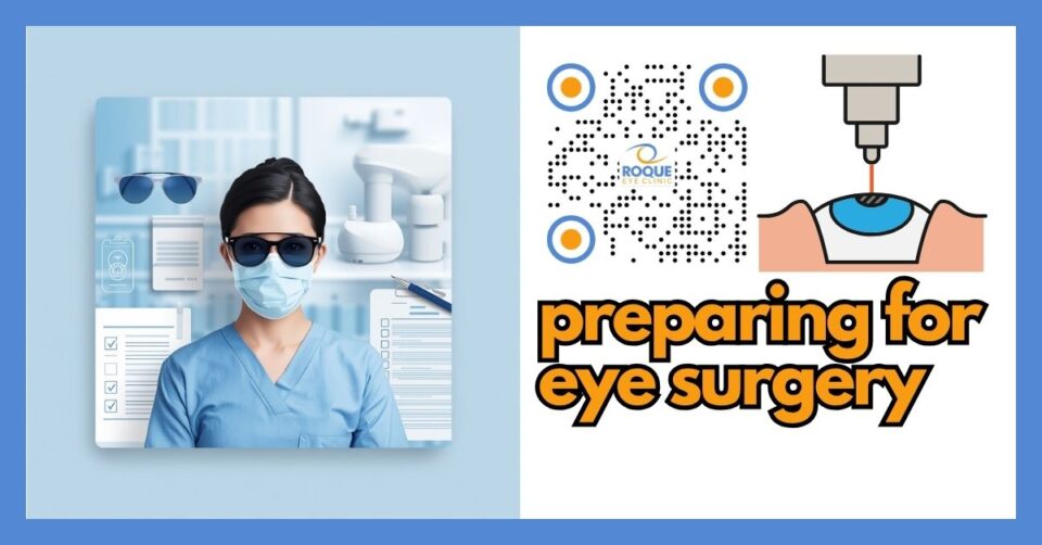
Neuroadaptation After Refractive Surgery
🧠 Quick Answer
Neuroadaptation after refractive surgery is the brain’s adjustment period after your eyes receive a new optical system. During this time, symptoms such as glare, halos, waxy vision, reduced contrast, imbalance between the eyes, or awareness of visual imperfections may gradually become less bothersome. Some patients adapt within weeks, while others need months, and some symptoms may persist if the problem is not only brain adaptation but also a true optical or ocular-surface issue.
Many patients assume vision recovery after refractive surgery is only about the cornea or lens healing. In reality, the brain also has work to do. Even when surgery is technically successful, the visual system may need time to interpret a new pattern of focus, blur, contrast, light scatter, or depth of field. That adjustment process is called neuroadaptation.
This matters because a patient may say, “My vision is sharp, but it still feels strange,” or “I see halos, but they bother me less now than they did last month.” Those statements often reflect neuroadaptation. At the same time, doctors must be careful not to blame every complaint on adaptation. Dry eye, residual refractive error, irregular astigmatism, decentered treatment, posterior capsule changes, or IOL-related issues can also cause persistent symptoms that need treatment rather than patience alone.
🧩 Focus: Neuroadaptation after laser and lens-based refractive surgery
👁 Goal: Explain why visual symptoms can feel different after surgery, what neuroadaptation means, how long it may take, and when symptoms need active evaluation
🛡 Evidence-Based: Preferred Practice Patterns • Standards of Care • Systematic Reviews • Meta-Analyses
REFRACTIVE SURGERY Knowledge Hub
Start with the complete guide:
🔬 Neuroadaptation After Refractive Surgery Anatomy Micro-Primer
- Cornea: The clear front window of the eye. Laser procedures reshape it, which can change how light enters the eye and how the brain experiences image quality.
- Lens or IOL: In lens-based surgery, the natural lens is replaced or supplemented. Multifocal, EDOF, or other optical designs can create new light-distribution patterns that the brain must learn to interpret.
- Retina: This is the light-sensitive tissue at the back of the eye. It sends the new image information to the brain after surgery.
- Visual cortex: This is the brain region that processes what the eyes see. Neuroadaptation mainly happens here, where the brain learns to pay more attention to useful information and less attention to unwanted optical noise.
📘 Neuroadaptation After Refractive Surgery Terminology Glossary
- Neuroadaptation: The brain’s gradual adjustment to a new optical system after surgery.
- Dysphotopsia: Unwanted visual phenomena such as glare, halos, arcs, shadows, or starbursts.
- Contrast sensitivity: The ability to distinguish an object from its background, especially in dim or low-contrast settings.
- Residual refractive error: Leftover nearsightedness, farsightedness, or astigmatism after surgery.
- Higher-order aberrations: Complex optical imperfections that can affect night vision, clarity, and quality of vision.
- Spectacle independence: Being less dependent on glasses for daily tasks after surgery.
Quick Navigation
Related Reading
Key Learning Points
- Neuroadaptation means the brain, not just the eye, is adjusting after refractive surgery.
- Symptoms such as glare, halos, waxy vision, reduced contrast, or imbalance between the eyes may improve with time.
- Laser procedures and lens-based procedures can both involve neuroadaptation, but the pattern may differ.
- Not all symptoms are “just in your brain”; dry eye, residual prescription, corneal irregularity, or IOL issues may need treatment.
- Good preoperative counseling helps patients tolerate the adjustment period more successfully.
What Neuroadaptation Is
Neuroadaptation is the visual system’s learning process after surgery changes the way light is focused or distributed in the eye. Before surgery, the brain becomes very used to a particular optical pattern, even if that pattern includes blur from refractive error or image distortion from spectacles, contact lenses, cataract, or an aging lens. After surgery, the optical system changes suddenly. The eyes may heal within days or weeks, but the brain may need longer to decide which parts of the new image to emphasize and which parts to ignore.
This is why some patients say, “I can see well, but the image still feels unnatural.” The eye may be physically fine, yet the visual brain has not fully settled into the new system. That is especially relevant when surgery increases depth of focus, changes binocular balance, or introduces new light phenomena that the patient never experienced before.
💡 Analogy
Neuroadaptation is like moving into a new home beside a busy road. At first, you hear every passing car. Over time, your brain stops treating the sound as important, and you notice it less. After refractive surgery, the brain may gradually learn to “tune down” halos, blur awareness, or minor optical distractions in a similar way.
Why It Happens After Refractive Surgery
Refractive surgery changes optics in a way that can be dramatic. After LASIK, PRK, or SMILE, the cornea may deliver a sharper but different visual signal. After ICL, refractive lens exchange, or premium IOL implantation, the eye may receive a new pattern of focus and light distribution. The brain must then decide how to process this signal efficiently.
Several mechanisms can contribute:
- New image geometry: The brain is receiving a pattern of focus that differs from the preoperative state.
- Change in binocular balance: If one eye heals faster, is targeted differently, or has different quality of vision, the brain may need time to coordinate both eyes comfortably.
- Awareness of dysphotopsia: Halos, glare, starbursts, or edge effects may initially capture the patient’s attention more strongly.
- Depth-of-focus trade-offs: Presbyopia-correcting strategies often improve range of vision but can alter contrast or night-quality perception.
- Expectation mismatch: The more a patient expects “perfect” vision instantly, the harder the adaptation phase may feel.
Common Symptoms During Neuroadaptation
Symptoms vary by procedure and by patient, but common complaints include:
- Halos around lights at night
- Glare or starbursts
- Ghosting or double contours
- Waxy, smeary, or “not crisp enough” vision
- Reduced contrast, especially in dim lighting
- Imbalance between distance and near perception
- Eye dominance confusion after monovision or blended-vision strategies
- Feeling that vision is technically good but still “strange” or “unfamiliar”
These symptoms do not automatically mean something went wrong. They may reflect early healing, ocular surface instability, or normal neural adjustment. The key question is whether symptoms are gradually improving, staying the same, or getting worse.
Laser Vision Correction Versus Lens-Based Surgery
After laser vision correction
After LASIK, PRK, TransPRK, or SMILE, the brain often adapts to a new clarity profile, especially if the patient previously depended heavily on glasses or contact lenses. Some symptoms are not purely neural. Dry eye, epithelial remodeling, flap or interface effects, and residual refractive error can all affect early quality of vision. Still, the patient’s awareness of glare, halos, and night symptoms may diminish with time as the brain becomes less bothered by them.
After lens-based surgery
Neuroadaptation is often discussed more in lens-based surgery, especially with multifocal and extended depth-of-focus IOLs. These optics may intentionally split light or alter the way focus is distributed across distances. The patient can gain greater spectacle independence, but some people initially notice rings, glare, halos, or reduced contrast more strongly. AAO discussions of dysphotopsia and multifocal/EDOF lenses note that symptoms often improve over weeks to months, though not all patients adapt equally well.
Why Some Patients Adapt Easily and Others Do Not
Neuroadaptation is not identical for everyone. Several factors matter:
- Personality and attention style: Patients who are highly detail-focused may remain more aware of subtle visual imperfections.
- Expectations: Patients seeking “zero awareness of any optical effect” may struggle more than patients prepared for temporary adaptation.
- Ocular surface quality: Dry eye can keep the image unstable, making adaptation harder.
- Residual refractive error: Even small leftover prescription can amplify dissatisfaction.
- Procedure type: Multifocal and presbyopia-correcting optics often demand more adaptation than monofocal optics.
- Binocular match: The brain adapts more easily when both eyes work together smoothly.
How Long Neuroadaptation May Take
There is no single exact timeline. Some patients feel much better within days or weeks. Others need several months. AAO discussions of postoperative dysphotopsia note that improvement often occurs around four to six weeks, but in some patients neuroadaptation may take up to a year. In multifocal or EDOF IOL patients, adaptation may be quicker in some and slower in others. A symptom that is steadily improving is usually more reassuring than one that is unchanged or worsening.
It is often helpful to think of the timeline in phases:
- Early phase: healing and inflammation dominate; vision may fluctuate a lot.
- Intermediate phase: the brain begins to ignore some unwanted visual noise.
- Late phase: symptoms may become much less noticeable, or persistent non-neural problems become easier to identify.
When It Is Not Just Neuroadaptation
This is one of the most important counseling points. Neuroadaptation is real, but it should never be used as a blanket explanation for every unhappy patient. Symptoms may persist because there is a treatable optical or medical issue. Examples include:
- Dry eye or meibomian gland dysfunction
- Residual refractive error or astigmatism
- Irregular corneal healing or haze
- Decentration or optical zone mismatch
- Pupil-related night-vision problems
- IOL malposition, dysphotopsia, or poor lens choice for the patient’s lifestyle
- Posterior capsular opacification in lens-based surgery
- Retinal or neurologic causes of visual dissatisfaction
If the image is unstable, adaptation becomes harder. In other words, the brain cannot learn a moving target very well. That is why treatment of dry eye, ocular surface disease, or residual refractive error may dramatically improve what initially looked like a “neuroadaptation problem.”
🚨 Emergency Warning
Seek urgent ophthalmic review if you have sudden major vision loss, a curtain over vision, flashes with many new floaters, severe pain, marked redness, or rapid worsening rather than gradual improvement. These are not typical neuroadaptation symptoms and may signal a retinal, inflammatory, pressure-related, or other urgent eye problem.
What May Help Neuroadaptation
Management depends on whether symptoms are primarily neural, optical, or mixed. Helpful steps may include:
- Time and reassurance: when the exam is reassuring and symptoms are trending better.
- Ocular surface treatment: lubrication, blink support, lid treatment, or anti-inflammatory therapy when needed.
- Residual error correction: temporary glasses, contact lenses, or enhancement planning when appropriate.
- Night-environment counseling: recognizing that night driving may be more challenging early on.
- Expectation coaching: reframing the goal from “perfect optics” to useful, functional, satisfying vision.
- Binocular adaptation support: especially for monovision, blended vision, or presbyopia strategies.
The Role of Expectations
Expectation management is one of the most powerful ways to improve patient satisfaction. A patient who understands before surgery that the brain may need time is less likely to panic in the early postoperative period. A patient who expects instant “HD vision” without any awareness of halos or contrast trade-offs is more likely to be distressed, even if the surgery is objectively successful.
This is why some surgeons say patients should aim for 20/happy, not necessarily “perfect vision in every lighting condition at every distance without any optical side effect.” That does not mean settling for poor outcomes. It means recognizing that some refractive solutions involve trade-offs, and patient success depends partly on adapting to those trade-offs.
How Doctors Evaluate a Patient Who Is Not Adapting Well
When symptoms remain bothersome, a careful review usually includes refraction, ocular surface evaluation, corneal assessment, pupil discussion, and if relevant, IOL centration and posterior segment review. The main goal is to separate:
- normal adaptation
- healing-related issues
- true optical problems that need treatment
Sometimes the best answer is reassurance and time. Sometimes the right answer is not patience but intervention, such as dry eye treatment, enhancement, lens exchange discussion, or a different corrective strategy.
Continue Reading
🏁 Take-Home Message
Neuroadaptation after refractive surgery is the brain’s adjustment to a new way of seeing. It can make early symptoms feel less noticeable over time, especially after premium optics or any procedure that changes the eye’s focusing system. But not every complaint is “just neuroadaptation.” If symptoms are severe, worsening, or not improving, the eye itself must be checked carefully for treatable causes.
FAQ
1) What does neuroadaptation mean after refractive surgery?
It means your brain is learning to process a new optical system after surgery. Even if the procedure is successful, your visual system may need time to feel comfortable with the new image quality.
2) How long does neuroadaptation usually take?
Some patients improve within weeks, while others need months. In some lens-based cases, adaptation may continue for a long time, and improvement can still occur later rather than immediately.
3) Are halos always a sign that something went wrong?
No. Halos can happen during normal healing and adaptation, especially at night. But persistent or severe halos should still be assessed to rule out dry eye, residual refractive error, pupil-related effects, or optical problems.
4) Is neuroadaptation more common with multifocal or EDOF lenses?
It is often discussed more with multifocal and EDOF lenses because these designs can change how light is distributed and can create more noticeable early visual phenomena that the brain must learn to handle.
5) Can dry eye make neuroadaptation harder?
Yes. If the tear film is unstable, the image reaching the brain is also unstable. That makes it harder for the visual system to settle comfortably.
6) What should I do if my vision still feels strange months after surgery?
You should have a careful postoperative evaluation. The cause may be adaptation, but it could also be a treatable issue such as residual prescription, ocular surface disease, corneal irregularity, or an IOL-related problem.
📚 References
- American Academy of Ophthalmology. Managing Dysphotopsias From Cataract Surgery. 2023.
- American Academy of Ophthalmology. New Lens, Same Brain: The Importance of Neuroadaptation. 2007.
- Rampat R, Gatinel D. Multifocal and Extended Depth-of-Focus Intraocular Lenses in 2020. Ophthalmology. 2021;128(11):e164-e185.
- U.S. Food and Drug Administration. When is LASIK not for me?
- U.S. Food and Drug Administration. LASIK Quality of Life Collaboration Project.
🤝 Roque Eye Clinic Patient Education Series
Dr. Manolette Roque | Dr. Barbara Roque
St. Luke’s Medical Center Global City | Asian Hospital Medical Center
Philippines
Medical Review: Roque Advisory Council
Last Updated: March 2026
Medical Disclaimer
This article is intended for educational purposes only and does not replace professional medical consultation.
