Key Learning Points
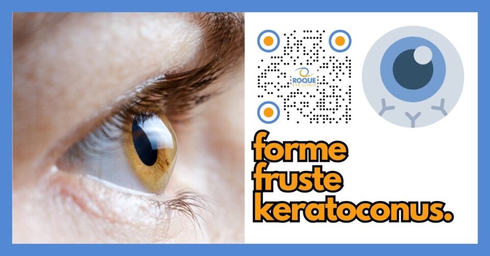
Forme fruste keratoconus (FFKC) is the “stealth‑mode” version of keratoconus—the cornea still looks normal at the slit lamp, but sophisticated imaging can already pick up early structural weakness.
Because FFKC can rapidly progress once it crosses the clinical threshold, early detection is critical—especially before laser vision‑correction surgery or in young patients with high eye‑rub habits.
Modern Scheimpflug, OCT‑based tomography, epithelial‑mapping, and artificial‑intelligence (AI) indices can spot FFKC with >95 % accuracy when used together.
Prompt corneal collagen cross‑linking (CXL) can “freeze” the disease and save vision, with comparable safety between epi‑off and newer accelerated or epi‑on protocols.
Rubbing control, allergy treatment, and good eye‑protection habits remain the best daily defenses against progression.
What Is Forme Fruste Keratoconus?
Forme fruste keratoconus (FFKC) literally means “an incomplete form of keratoconus.” Imagine the cornea—the clear front window of the eye—as a perfectly round drumhead. In classic keratoconus, that drumhead slowly thins and sags into a cone, scattering incoming light and blurring vision. In FFKC, the drumhead has just begun to lose its even tension. To the naked eye it still looks flat, but sensitive instruments can already detect subtle dips and stretches.
The Spectrum of Disease
| Stage | What Your Doctor Sees | What You May Notice |
|---|---|---|
| Normal cornea | Symmetrical thickness and shape | Clear vision |
| FFKC (subclinical) | Normal slit‑lamp exam; suspicious tomography/AI flags | None or mild ghosting/glare in dim light |
| Clinical keratoconus | Cone on topography, visible thinning, possible scars | Blurred, distorted vision, frequent glasses changes |
Why Does FFKC Happen?
Researchers believe FFKC arises from a perfect storm of factors:
Genetics – Up to 1 in 5 patients report an affected relative. Specific gene variants in collagen and extracellular‑matrix pathways increase risk.
Eye Rubbing & Allergy – Chronic mechanical trauma plus inflammatory mediators weaken corneal collagen cross‑links.
Hormonal & Environmental Stress – Puberty, pregnancy, ultraviolet light, and oxidative stress may tip a borderline cornea into ectasia.
Symptoms: Why FFKC Is Usually Silent
Most patients have no symptoms at all. When symptoms do appear they are subtle:
Blurry distance vision that “comes and goes”
Ghost images or halos around lights at night
Increased sensitivity to glare or eye strain
Because these signs overlap with simple astigmatism, many patients are only diagnosed when they seek laser vision correction or a contact‑lens refit.
How We Detect FFKC in 2025
Advanced imaging is our stethoscope. We combine four pillars:
| Pillar | What It Measures | Early Red‑Flag for FFKC |
|---|---|---|
| Scheimpflug tomography | 3‑D corneal shape/thickness (Pentacam, Sirius) | Skewed “butterfly” curvature, I‑S asymmetry |
| Anterior‑segment OCT | High‑resolution layer thickness | Localized epithelial thinning over steep zones |
| Biomechanical analyzers | How the cornea flexes to air‑puff | Reduced corneal stiffness indices |
| AI composite indices | Machine‑learning risk score | >0.3 Abbruzzese or BAD‑D index |
Meta‑analysis shows pooled sensitivity 98.6 % and specificity 98.3 % for detecting subclinical keratoconus when multiple maps are combined.
Treatment Options
1. Observation with Lifestyle Control
Stop eye rubbing—use cold compresses or antihistamine drops for itch.
Wear UV‑blocking sunglasses.
Follow 6‑month imaging schedule.
2. Corneal Collagen Cross‑Linking (CXL)
CXL uses riboflavin (vitamin B2) and ultraviolet‑A light to stiffen corneal collagen, much like adding extra rebar to concrete.
| Protocol | Typical Time | Key Points |
|---|---|---|
| Epi‑off Dresden (standard) | 30 min UV | Gold standard flattening, more discomfort |
| Accelerated epi‑off | 10 min UV | Similar effect, less chair time |
| Transepithelial (epi‑on) | 30–40 min UV | Less pain, slightly less stiffening; good for borderline corneas |
| Pulsed/Topography‑guided | Variable | Customized energy for thin or irregular corneas |
3. Specialty Contact Lenses
Soft toric, hybrid, or scleral lenses improve optics without surgery.
4. Future Therapies
Gene editing, customized UV patterns, and riboflavin nano‑carriers are under study.
Prevention & Daily Care
Think of your cornea as a camping tent. Yanking the tent pole (eye rubbing) or constant wind (UV light) loosens the fabric. Protect it by:
Hands‑off rule—rub around the eye, not on the cornea.
Allergy control—daily antihistamine drops when pollen counts rise.
UV defense—broad‑brimmed hat + 100 % UV‑blocking sunglasses.
Balanced screen time—every 20 minutes, look 20 feet away for 20 seconds (20‑20‑20 rule).
Post‑Treatment Healing Steps
First 48 hours – Use antibiotic + steroid drops as prescribed. Expect gritty sensation.
Week 1 – Avoid swimming and eye makeup. Wear the provided bandage contact lens (if epi‑off).
Week 4 – Gradually resume contact‑lens wear once cleared.
Month 3 & 6 – Repeat tomography to confirm corneal flattening.
Lifelong – Annual scans; continue rub‑free habits.
Analogy
Picture your cornea like a trampoline mat. When the springs (collagen fibers) loosen unevenly, the center sags. Cross‑linking is like re‑tensioning those springs with extra zip‑ties so the mat stays flat and you can keep “seeing” the world clearly.
Frequently Asked Questions
Is FFKC the same as keratoconus?
No. It is the earliest, subclinical stage—think of a seed rather than a tree.Will it always progress?
About 30‑50 % progress within 5 years, faster in teenagers and habitual eye rubbers.Can glasses fix my vision?
Yes, in the very early stage. If distortions increase, specialty contact lenses offer clearer optics.Is cross‑linking painful?
Epi‑off feels like a scratchy eye for 2–3 days; epi‑on is milder.How long is recovery?
Functional vision usually returns in 1–2 weeks, with steady corneal flattening over 6–12 months.Can I have LASIK later?
Usually no—LASIK removes tissue and can destabilize the cornea. Other refractive options (ICL, PRK after CXL) may be considered case‑by‑case.Will insurance cover CXL?
Coverage varies; bring documentation of progression and discuss with your provider.Is there any medicine to stop FFKC?
No drops can currently stiffen collagen like CXL; antioxidant supplements are still investigational.What happens if I ignore it?
Advanced keratoconus may require corneal transplantation with longer recovery and higher cost.Can both eyes be treated on the same day?
Many surgeons treat one eye at a time to monitor healing, but same‑day bilateral CXL is safe in selected cases.
Take‑Home Message
Early detection and timely cross‑linking transform FFKC from a vision‑threatening surprise into a manageable condition. Protect your eyes, stop the rub, and partner with your cornea specialist for lifelong clear vision.
Bibliography Lists
1. Definitions & Epidemiology
Henriquez MA, Hadid M, Izquierdo L Jr. A systematic review of subclinical keratoconus and forme fruste keratoconus. J Refract Surg. 2020;36(4):270‑279.
Smadja D, et al. New ways to detect keratoconus. Rev Ophthalmol. 2014;21(9):34‑40.
Klyce SD. Catching keratoconus: making the tough calls. Rev Ophthalmol. 2010;17(7):42‑48.
2. Diagnostic Technology
Hashemi H, Doroodgar F, Niazi S, et al. Artificial intelligence for detecting keratoconus: systematic review and meta‑analysis. Graefes Arch Clin Exp Ophthalmol. 2024;262(4):1017‑1039.
Cao K, Verspoor K, Sahebjada S, Baird PN. Accuracy of machine‑learning detection of keratoconus. J Clin Med. 2022;11(3):478.
Talpan C, et al. Universal architecture of corneal segmental tomography biomarkers. Br J Ophthalmol. 2023;107(6):848‑856.
3. Risk Factors & Genetics
Kandel H, et al. Non‑genetic risk factors for keratoconus: systematic review. Eye Contact Lens. 2023;49(2):78‑90.
NEI News Release. Potential causes of keratoconus uncovered. Natl Eye Inst. 2023.
Pur DR, et al. AI analysis of biofluid markers in corneal disease: systematic review. Eye (Lond). 2023;37(10):2007‑2019.
4. Cross‑Linking Protocols
López‑Marín L, et al. Epithelium‑on vs. epithelium‑off cross‑linking for keratoconus: meta‑analysis. Cornea. 2023;42(11):1395‑1405.
Chen Q, et al. Accelerated vs. conventional CXL: randomized controlled trial meta‑analysis. Ophthalmology. 2024;131(2):142‑151.
Ilhan‑Sahinoz A, et al. Pulsed corneal cross‑linking in keratoconus: systematic review. Eye Vis (Lond). 2024;11:25.
5. Long‑Term Outcomes & Future Directions
Gupta A, et al. Long‑term safety and efficacy of corneal collagen cross‑linking: systematic review. Ocul Surf. 2023;28:320‑334.
O’Brart D, et al. Epithelial & stromal effects of cross‑linking: comprehensive review. Prog Retin Eye Res. 2023;93:101119.
Tang C, et al. Posterior‑to‑anterior corneal curvature ratio distribution in myopes. Front Med. 2021;8:724674.
BOOK AN APPOINTMENT
It takes less than 5 minutes to complete your online booking. Alternatively, you may call our BGC Clinic, or our Alabang Clinic for assistance.
